
Physiology of freediving
.jpg "A freediver descending on a breath hold. Depth freediving combines prolonged apnea, exercise, immersion and increasing hydrostatic pressure.") Physiology of freediving, also called breath-hold diving physiology, concerns the physiological responses, adaptations and pathological effects associated with freediving and prolonged voluntary apnea. Freediving combines several physiological stresses: interruption of pulmonary ventilation, immersion, exercise, and, during depth diving, large changes in ambient pressure. These produce progressive hypoxemia, hypercapnia and acid–base disturbance, while hydrostatic pressure alters lung volume, pulmonary blood volume and the partial pressures of respiratory gases.
Physiology of freediving, also called breath-hold diving physiology, concerns the physiological responses, adaptations and pathological effects associated with freediving and prolonged voluntary apnea. Freediving combines several physiological stresses: interruption of pulmonary ventilation, immersion, exercise, and, during depth diving, large changes in ambient pressure. These produce progressive hypoxemia, hypercapnia and acid–base disturbance, while hydrostatic pressure alters lung volume, pulmonary blood volume and the partial pressures of respiratory gases.
Humans respond to apnea and immersion with the diving response, which includes bradycardia, peripheral vasoconstriction and redistribution of blood flow towards organs with high oxygen requirements. During deep dives, compression of pulmonary gas is accompanied by an increase in central and pulmonary blood volume, commonly called the blood shift. These responses extend tolerance of apnea and pressure but do not prevent progressive depletion of oxygen stores or the development of pulmonary, cardiovascular and neurological limits.
At physiological extremes, mechanisms that are normally protective can become inadequate or contribute to pathology. Important adverse phenomena include hypoxic loss of motor control, freediving blackout, pulmonary barotrauma of descent ("lung squeeze"), middle-ear and sinus barotrauma, cardiac rhythm disturbances, and, in sufficiently deep or repetitive diving, decompression sickness.
Terminology
Terminology in freediving overlaps with, but does not always match, physiological and medical usage.
A prolonged voluntary apnea is commonly divided into an easy-going phase and a struggle phase. The transition, or physiological breakpoint, is marked by the onset of involuntary breathing movements (IBMs), commonly called contractions by freedivers.
Hypoxic neurological impairment without complete loss of consciousness is termed loss of motor control (LMC); the associated tremulous or jerking movements are commonly called a "samba".
Terminology for hypoxic blackout is inconsistent. The term shallow-water blackout has been applied both to hypoxic blackout during shallow or approximately constant-pressure breath holding, particularly following hyperventilation, and to blackout during the final part of ascent from a deep breath-hold dive. In the latter, falling ambient pressure causes a rapid decline in alveolar and arterial oxygen partial pressure. This mechanism is more unambiguously termed ascent blackout; deep-water blackout has also been used for it, although the term can be misleading because loss of consciousness commonly occurs near the surface.
Glossopharyngeal insufflation is commonly called lung packing or packing. Redistribution of blood into the thoracic and pulmonary circulation during immersion and lung compression is commonly termed the blood shift. Pulmonary injury associated with extreme compression during descent is commonly called lung squeeze.
Physiology of prolonged apnea
Easy-going phase and physiological breakpoint
 Maximal voluntary apnea is commonly divided physiologically into an easy-going phase followed by a struggle phase. During the easy-going phase, ventilation is voluntarily suppressed and overt involuntary respiratory muscle activity is absent or limited, although PaCO2 is progressively rising and PaO2 is falling. The duration of this phase depends on factors including initial blood gases, lung volume, metabolic rate and individual chemosensitivity to oxygen and carbon dioxide.
The transition between the phases is termed the physiological breakpoint and is associated with the appearance of involuntary breathing movements (IBMs), commonly called contractions by freedivers. These movements are generated as chemical and neural respiratory drive becomes sufficiently strong to produce involuntary activity of the diaphragm and other respiratory muscles despite continued voluntary closure of the airway.
The onset of IBMs does not appear to be determined by a single oxygen or carbon dioxide value. In an experiment that varied inspired oxygen and carbon dioxide and obtained arterial samples at IBM onset, Breskovic et al. identified a possible PaCO2 threshold of approximately 6.5 ± 0.5 kPa (48.8 ± 3.8 mmHg). No equivalent single PaO2 threshold was found, and the results indicated an interaction between oxygen and carbon dioxide in determining the breakpoint. The value is therefore an experimental estimate rather than a universal threshold for the first "contraction".
Struggle phase and involuntary breathing movements
During the struggle phase, repeated IBMs generate fluctuations in intrathoracic and abdominal pressure. The movements may influence venous return, stroke volume and cerebral circulation while the diver becomes progressively more hypoxemic and hypercapnic. They also impose repeated mechanical work on the diaphragm and other respiratory muscles, and respiratory-muscle fatigue after prolonged or repeated maximal apnea has been proposed as an additional performance limitation.
A 2026 multimodal case study of a world-champion freediver illustrates the magnitude of this response. During a maximal dry static apnea lasting 6 min 7 s, the first 2 min 20 s comprised the easy-going phase and the remaining 3 min 47 s the struggle phase; electromyography recorded 48 involuntary breathing movements. End-tidal carbon dioxide rose from 21 mmHg after preparatory breathing to 65 mmHg at termination, while oxygen saturation fell from 97% to 73%. Muscle oxygenation declined substantially, whereas cerebral oxygenation was relatively preserved until late in the breath hold. Because end-tidal gases were measured before and after the apnea rather than at the first IBM, the terminal PETCO2 of 65 mmHg should not be interpreted as the threshold for contraction onset.
Human diving response
 The human diving response, often called the mammalian diving reflex, is an integrated autonomic response to apnea that is enhanced by facial immersion. Its characteristic cardiovascular components include bradycardia, peripheral vasoconstriction and redistribution of cardiac output. These responses reduce perfusion of less immediately oxygen-sensitive peripheral tissues and contribute to maintenance of oxygen delivery to the brain and heart.
Bradycardia reduces myocardial oxygen demand, while sympathetic vasoconstriction increases systemic vascular resistance and restricts blood flow to limbs and other peripheral tissues. The response is dynamic rather than uniform: exercise, water temperature, emotional state, lung volume and the severity of hypoxia and hypercapnia all modify its magnitude.
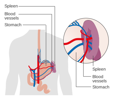
Cerebral blood flow increases substantially during severe apnea, partly in response to hypercapnia and falling arterial oxygen content. This helps preserve cerebral oxygen delivery despite progressive systemic hypoxemia. Contraction of the spleen during repeated or prolonged apnea can also transiently increase circulating erythrocyte concentration, thereby increasing blood oxygen-carrying capacity.
The simultaneous activation of sympathetic and parasympathetic pathways can also create electrophysiological instability. Marked sinus bradycardia, ectopic beats and other rhythm disturbances have been observed in breath-hold divers, particularly during severe apnea or deep diving. The clinical significance varies, and arrhythmia is not considered the mechanism of most freediving blackouts, which are primarily hypoxic.
Lung volumes and pulmonary mechanics
Lung volumes
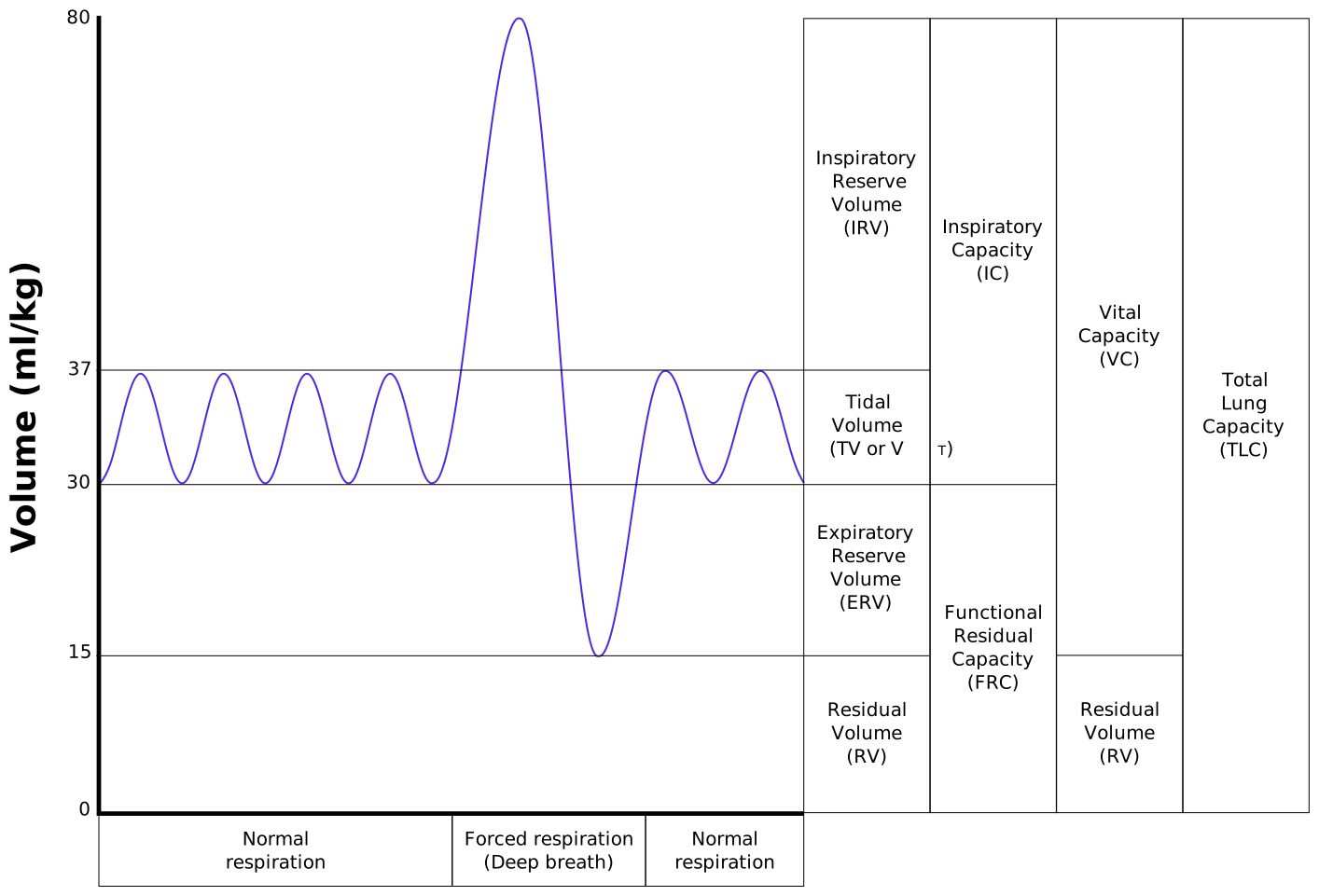 Several conventional lung volumes are important in freediving physiology. Total lung capacity (TLC) is the volume in the lungs after a maximal inspiration, while residual volume (RV) is the gas remaining after maximal expiration. Vital capacity is the volume between these limits. Functional residual capacity (FRC) is the volume remaining at the end of an ordinary passive expiration. These values determine both the initial pulmonary oxygen store and the degree to which the lungs can be compressed during descent.
Immersion itself changes lung mechanics before substantial depth-related compression occurs. In one study of eight trained divers and eight non-divers, FRC was approximately 30.8–34.8% lower while seated in water and 20.3–20.9% lower while prone in water than while seated in air. The change was principally mechanical and occurred in both trained and untrained participants.
Hydrostatic compression and residual-volume-equivalent depth
According to Boyle's law, increasing ambient pressure during descent reduces pulmonary gas volume approximately in inverse proportion to absolute pressure, although the human thorax and pulmonary circulation make the real situation more complex than a closed flexible gas container. Early models therefore predicted a depth at which the compressed lung would reach the diver's surface RV and beyond which further descent would be mechanically impossible or injurious.
In modern elite freedivers, dives substantially beyond such a simple RV-equivalent depth are possible. The surface RV is not a fixed minimum volume for the pressurised thorax: blood redistribution, deformation of the chest wall and diaphragm, regional airway closure and other mechanical changes allow total intrathoracic gas volume to fall below the value measured as RV at the surface.
The transition is nevertheless physiologically important. In a study of 25 divers performing 66 dives between 18 and 117 m, dives calculated to compress the lungs to or below the diver's surface RV were associated with greater transient post-dive impairment of pulmonary gas exchange. Nine minutes after diving, the estimated oxygen deficit was 24 ± 25 mmHg after dives reaching ≤RV compared with 5 ± 8 mmHg after dives not exceeding the RV-equivalent depth.
Thoracic blood shift
As hydrostatic pressure compresses the lungs, blood is redistributed from the peripheral venous circulation into the thorax and pulmonary circulation. This thoracic blood shift—usually shortened to blood shift in freediving terminology—increases pulmonary vascular blood volume and occupies part of the intrathoracic space surrendered by the compressed gas.
The blood shift is therefore an important reason why the surface RV does not constitute a rigid depth limit. It should not, however, be understood as simply preventing all lung collapse. Regional small-airway closure and alveolar collapse can still occur, particularly at extreme compression, and pulmonary gas exchange becomes less efficient in some deep divers. At high enough stress, pulmonary vascular engorgement and pressure gradients may contribute to capillary leakage, edema and hemorrhage.
Glossopharyngeal insufflation
Glossopharyngeal insufflation (GI), commonly called lung packing or packing, is used by some freedivers to increase pulmonary gas volume above that achieved by a conventional maximal inspiration. Sequential glossopharyngeal movements transfer additional gas into the lungs, increasing the initial pulmonary gas and oxygen reservoir and altering the depth at which a given degree of compression is reached.
The physiological effects are not limited to lung volume. The elevated intrathoracic pressure generated by substantial packing can impede venous return to the heart and transiently reduce cardiac output, arterial pressure and cerebral blood flow. In a small study of two world-class competitors, the diver who increased pulmonary volume by 22% showed greater initial hemodynamic disturbance than the diver whose volume increased by 10%.
Extremely high transpulmonary pressures produced by GI have also been associated with pulmonary barotrauma. Consequently, packing represents both a physiological means of increasing starting lung volume and a source of additional cardiopulmonary stress.
Respiratory gases and acid–base physiology
Oxygen stores and hypoxemia
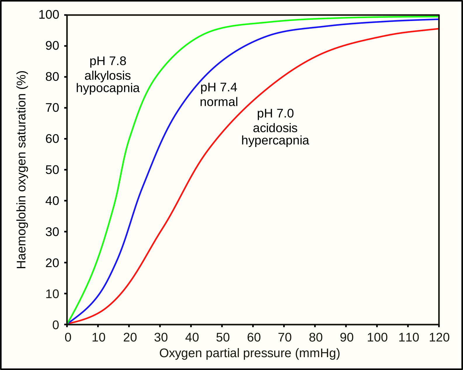 Oxygen available during a breath hold is distributed between pulmonary gas, hemoglobin-bound oxygen in the blood, and tissue stores including myoglobin in skeletal muscle. Pulmonary oxygen constitutes a large immediately available store at the beginning of apnea, but the relative importance of blood and tissue stores increases as the lungs become compressed and oxygen is transferred into the circulation.
Progressive oxygen consumption reduces arterial oxygen tension and eventually arterial oxygen saturation. Cerebral function depends on oxygen delivery rather than arterial oxygen tension alone, so consciousness is influenced by hemoglobin concentration, cerebral blood flow, cardiac output, carbon dioxide tension and individual cerebrovascular responses as well as PaO2. For this reason, no single PaO2 or pulse-oximeter saturation defines a universal threshold for loss of motor control or blackout.
Carbon dioxide and respiratory drive
Carbon dioxide produced by metabolism accumulates throughout apnea. The resulting hypercapnia contributes strongly to respiratory drive through central and peripheral chemoreceptors and is an important determinant of the transition from the easy-going phase to the struggle phase. Hypercapnia also causes cerebral vasodilation, which can support cerebral oxygen delivery even while systemic oxygenation declines.
Preparatory hyperventilation lowers PaCO2 before apnea. This can delay the rise of carbon dioxide to levels that produce strong respiratory drive and therefore prolong the period before the physiological breakpoint. It does not proportionately increase the body's oxygen store and can allow severe hypoxemia to develop before respiratory discomfort forces termination of the breath hold, which is one mechanism of constant-pressure hypoxic blackout.
Arterial gases at depth
Hydrostatic pressure makes the blood-gas trajectory of depth diving different from that of static apnea. Compression raises alveolar gas partial pressures during descent, causing arterial PaO2 and PaCO2 to rise even while oxygen is being consumed and carbon dioxide produced. At greater depth, once the largest proportional changes in lung volume have already occurred, metabolism increasingly determines the direction of the arterial gas changes.
A 2026 study directly sampled radial arterial blood in three elite freedivers during dives to 20, 40, 60 and 80 m. Following marked pre-dive hypocapnia, mean PaCO2 was 5.61 kPa (42.1 mmHg) at 40 m, 5.54 kPa (41.6 mmHg) at 60 m, and 6.9 kPa (51.5 mmHg) at 80 m. Mean PaO2 showed its largest compression-associated increase at 40 m, reaching approximately 29.3 kPa (220 mmHg).
The investigators interpreted the measurements as showing a compression-related arterial-gas effect that was greatest while lung volume was undergoing its largest proportional reduction. Beyond this region, continuing metabolism became increasingly evident: arterial oxygen fell while carbon dioxide accumulated. In these divers, the largest compression-associated effect occurred at approximately 40 m, around the depth at which simple lung-volume calculations placed the lungs near their surface RV.
The 2026 study involved only three divers and does not establish a universal carbon dioxide threshold for impairment at depth. It does, however, directly demonstrate that significant hypercapnia can coexist with pressure-induced hyperoxia during deep descent. Hypercapnia has been proposed as one contributor to cognitive or narcosis-like effects during very deep breath-hold dives, alongside the high partial pressure of nitrogen, but the relative contribution of each gas remains incompletely resolved.
Hyperoxic apnea and cerebral metabolism
Experiments using oxygen prebreathing allow very prolonged static apnea to be studied while delaying the development of hypoxemia. Such experiments can help separate effects of hypercapnia from those of oxygen deprivation and are distinct from ordinary air-breathing freediving.
Bain et al. found that cerebral oxidative metabolism decreased during extreme apnea and that severe hypercapnia accounted for part of this reduction. In a later study of ten ultra-elite apneists, protocols producing severe hypoxemic hypercapnia and prolonged hyperoxic hypercapnia produced different patterns of cerebral blood flow, oxidative metabolism and oxidative–nitrosative stress, illustrating that high carbon dioxide itself has substantial cerebrovascular and metabolic effects rather than acting only as a signal to breathe.
These acute changes in cerebral oxidative metabolism are distinct from proposed long-term adaptations of skeletal-muscle mitochondria in trained breath-hold divers.
Static and dynamic apnea
Static apnea provides an experimental model of prolonged breath holding with minimal locomotor oxygen demand. The principal changing variables are therefore the progressive disturbance of blood gases, autonomic cardiovascular responses, respiratory drive and the mechanical effects of IBMs. Static apnea has consequently been widely used to study the physiological breakpoint, cerebral oxygenation and the limits of voluntary apnea. 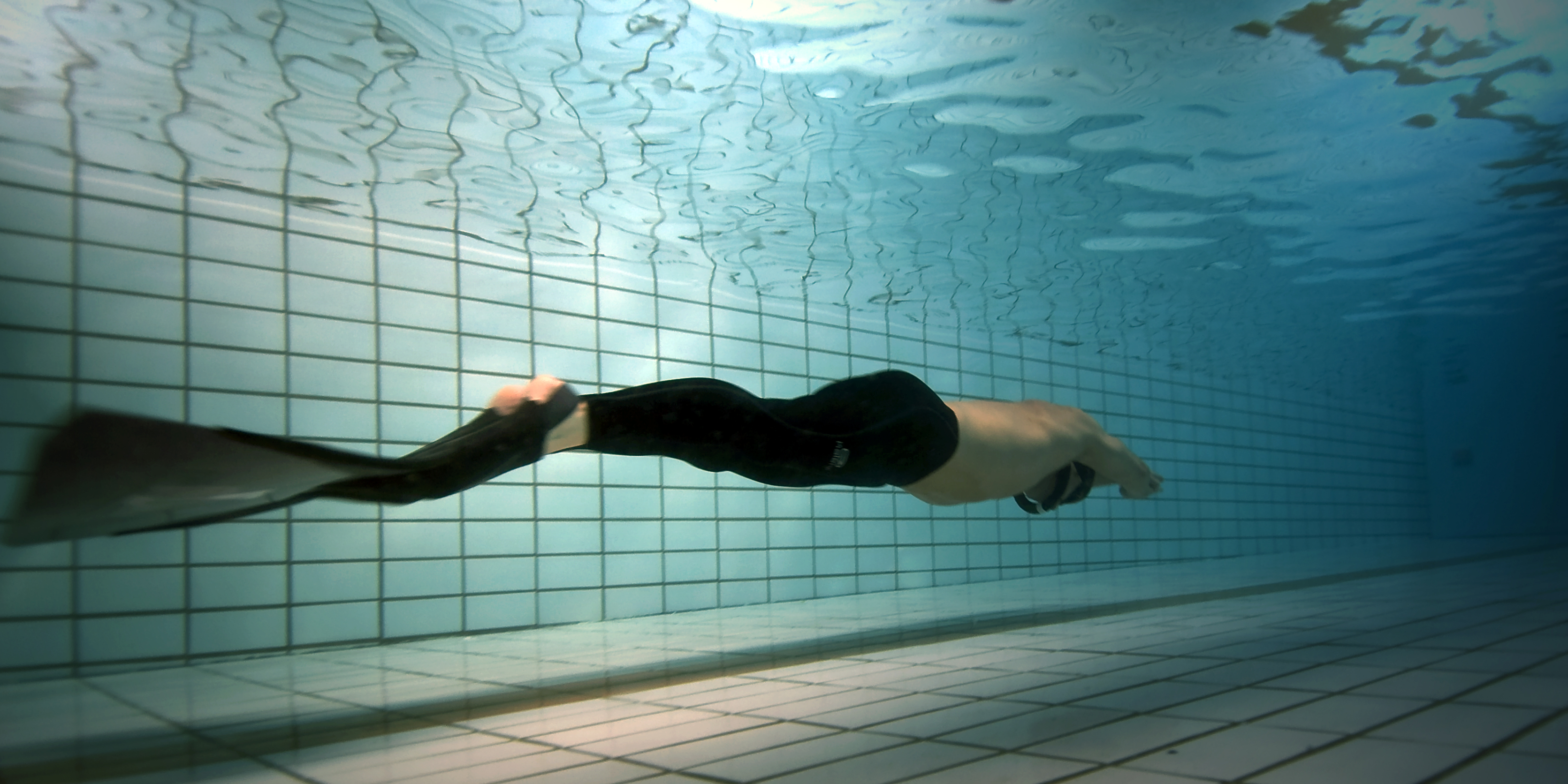 Dynamic apnea adds muscular work. Contracting skeletal muscle increases oxygen consumption and carbon dioxide production and can accelerate depletion of the oxygen reserve. Peripheral vasoconstriction produced by the diving response competes with the metabolic requirement for active-muscle blood flow; the balance changes with exercise intensity and training status.
The distinction is important when interpreting measurements. A breath-hold duration that is well tolerated during static apnea may produce substantially greater hypoxemia when the same duration includes swimming, while a depth dive adds pressure-dependent changes in gas partial pressures and lung mechanics that are absent in the pool.
Physiology of descent and maximum depth
During descent, increasing ambient pressure compresses pulmonary gas and raises its partial pressures. The diver consequently experiences a combination unusual in terrestrial physiology: progressive apnea with continuing metabolic oxygen consumption can coexist temporarily with rising pulmonary and arterial oxygen partial pressure caused by compression.
At the same time, lung volume falls, the diaphragm moves cranially, thoracic blood volume increases and small-airway closure may develop. These changes become especially important around and below the surface RV-equivalent depth. Gas exchange may continue despite very small lung volumes, but ventilation–perfusion inequality and diffusion limitations can increase.
Nitrogen partial pressure also rises with depth. Although the diver does not breathe continuously from a high-pressure gas supply, nitrogen already contained in the lungs can diffuse into blood and tissues while pulmonary gas exchange persists. At sufficient depths, high nitrogen partial pressures may contribute to nitrogen narcosis or narcosis-like symptoms. Concurrent hypercapnia may modify these effects.
Pressure equalisation of air spaces
Middle ear
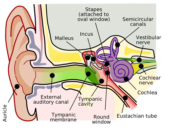 The gas-filled middle ear is connected to the nasopharynx by the Eustachian tube. During descent, external pressure rises while middle-ear gas is compressed. Without transfer of gas into the middle ear, an increasing pressure gradient develops across the tympanic membrane, causing discomfort and eventually middle-ear barotrauma.
Freedivers use pressure-equalisation manoeuvres including the Valsalva and Frenzel manoeuvres to generate a nasopharyngeal pressure sufficient to open the Eustachian tube. Their physiological importance increases with depth because progressive thoracic compression reduces the amount of readily transferable pulmonary gas. In a prospective study of 16 freedivers performing 317 dives to an average depth of 13.3 m freshwater, pressure-related Teed grade 1 or 2 changes were detected in 48% of examined ears; greater changes were associated with less diving experience, increasing depth and repeated exposure.
At greater depths, some freedivers use the mouthfill technique, in which gas transferred from the lungs earlier in descent is retained in the oral and pharyngeal space and later used to generate nasopharyngeal pressure when further transfer from the compressed thorax becomes difficult. In physiological terms, the technique extends the availability of an upper-airway gas reservoir for middle-ear equalisation. Published physiological research specifically examining mouthfill mechanics remains limited compared with the literature on other equalization techniques.
Paranasal sinuses
The paranasal sinuses are also gas-filled spaces, but they normally equalise passively through their ostia rather than requiring active opening comparable to the Eustachian tube. Obstruction of an ostium can prevent equilibration, creating a pressure gradient across the sinus mucosa and producing sinus barotrauma.
Equalisation therefore represents a physiological and anatomical depth constraint independent of oxygen stores. A diver may remain metabolically capable of further descent while being limited by the ability to maintain tolerable pressure gradients across the middle ear, sinuses or other gas-containing structures.
Physiology of ascent
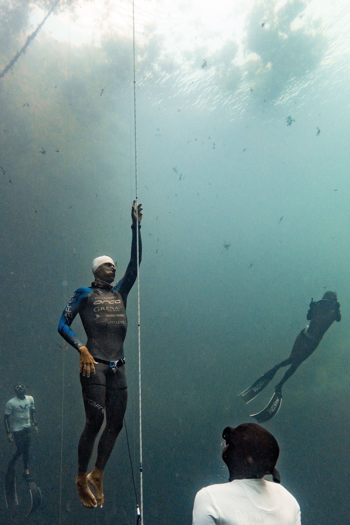 Ascent reverses hydrostatic compression. Pulmonary gas expands, central blood redistribution decreases and the partial pressures of gases in the lungs fall. For oxygen, this occurs after a substantial fraction of the original oxygen store has already been consumed, creating a characteristic risk of rapidly worsening hypoxemia during the final part of a depth dive.
The proportional pressure change becomes especially large near the surface. Between 10 m seawater and the surface, ambient pressure falls from approximately two atmospheres absolute to one atmosphere. All else being equal, this approximately halves the pressure contribution to alveolar gas partial pressures. Oxygen can therefore remain adequate for consciousness at depth and subsequently fall to critically low levels during ascent.
Field measurements support an independent effect of depth on post-dive hypoxemia. In a 2023 study of competitive freedivers, dives averaging 53 ± 14 m produced a minimum pulse-oximeter saturation of 58 ± 17%, compared with 74 ± 17% after dives averaging 17 ± 4 m, despite similar dive durations. Proposed contributors included the rapid fall in alveolar oxygen pressure on ascent, greater work and oxygen consumption, and impaired pulmonary gas exchange in some deep dives.
Hypoxic neurological impairment and blackout
As cerebral oxygen delivery declines, neurological function may deteriorate before complete loss of consciousness. Manifestations range from subtle impairment of coordination or judgement to hypoxic loss of motor control (LMC) and ultimately blackout. Because cerebral perfusion and oxygen extraction vary between individuals and are strongly affected by carbon dioxide, there is no universally accepted arterial oxygen tension or saturation at which these events invariably occur.
Loss of motor control
Loss of motor control is the term commonly used in competitive freediving for transient hypoxic neurological dysfunction in which consciousness is at least partly retained but purposeful motor control is impaired. Freedivers commonly call the characteristic tremulous or jerking presentation a "samba". Reported manifestations include loss of postural control, confusion, difficulty speaking, spasmodic movements and impaired execution of voluntary actions.
Competition observations illustrate that LMC and complete blackout are distinct outcomes. In data analysed by Lindholm from competitions in 2002–2004, LMC occurred in 9.6% of 355 static-apnea performances and 6.1% of 344 constant-weight performances, while loss of consciousness occurred in 1.1% and 6.1%, respectively. These figures describe particular elite competition samples and should not be interpreted as general incidence rates for recreational freediving.
Constant-pressure hypoxic blackout
Constant-pressure hypoxic blackout describes loss of consciousness during a breath hold in which ambient pressure changes little, as in static apnea or pool dynamic apnea. Oxygen is progressively consumed until cerebral oxygen delivery becomes inadequate. Hyperventilation can increase risk by lowering the initial PaCO2 and delaying carbon-dioxide-mediated respiratory drive without comparably increasing the oxygen reserve.
This mechanism is frequently described in common usage as shallow-water blackout. The term has been applied inconsistently, however, and contemporary medical sources increasingly favour mechanistic terms such as hypoxic blackout.
Ascent-induced hypoxic blackout
Ascent-induced hypoxic blackout occurs after a depth dive when declining ambient pressure causes pulmonary and arterial oxygen partial pressures to fall during ascent. It is therefore mechanistically distinct from constant-pressure blackout even though the final cause of unconsciousness in both is cerebral hypoxia.
The common term deep-water blackout is potentially misleading because loss of consciousness often occurs during the final metres of ascent or immediately after surfacing rather than at maximum depth. Conversely, the expression shallow-water blackout has historically also been applied to blackout during the shallow final portion of ascent. For clarity, physiological literature increasingly distinguishes blackout by mechanism—constant-pressure hypoxic blackout versus ascent-induced hypoxic blackout—rather than solely by the depth at which consciousness is lost.
Pathophysiology
Pulmonary barotrauma of descent
Extreme hydrostatic lung compression can produce pulmonary symptoms collectively described by freedivers as lung squeeze. Clinical and physiological descriptions include chest discomfort, cough, hemoptysis, pulmonary edema, pulmonary hemorrhage and transient impairment of pulmonary gas exchange. The underlying mechanisms are thought to include very low lung volumes, large pressure gradients and high pulmonary capillary blood volume and pressure.
The thoracic blood shift is therefore both protective and potentially stress-producing. By increasing central vascular volume it allows the thorax to accommodate compression below the surface RV predicted by simple gas-volume models, but extreme vascular engorgement can increase pulmonary capillary stress. Regional airway closure and atelectasis may further impair gas exchange.
Glossopharyngeal insufflation creates a different pressure exposure at the beginning of the dive: instead of very low intrathoracic gas volumes, packing can generate abnormally high lung volumes and transpulmonary pressures. Pulmonary injury has been reported after forceful GI, illustrating that both extremes of lung volume can produce pathology.
Inert-gas uptake and decompression illness
Although a breath-hold diver carries only the gas contained in the lungs at the start of the dive, high ambient pressure substantially raises alveolar nitrogen partial pressure. As long as pulmonary gas exchange continues, nitrogen can enter the blood and tissues. With repetitive deep dives or a sufficiently long and deep single exposure, subsequent ascent can produce tissue supersaturation and bubble formation.
A 2023 systematic review identified 44 reported incidents of decompression illness across 17 publications involving breath-hold divers. Both decompression sickness caused by inert-gas supersaturation and mechanisms compatible with arterial gas embolism have been proposed in individual cases. Repetitive-diving neurological illness described historically in Polynesian breath-hold divers is known as Taravana.
Extreme single breath-hold dives can also produce clinically significant decompression illness. A prominent example is Herbert Nitsch, who developed severe neurological decompression sickness after an extreme no-limits dive in 2012; the case has been cited in physiological reviews as evidence that a single unusually deep and prolonged breath-hold exposure can produce substantial inert-gas loading.
Training and physiological adaptation
Repeated apnea training is associated with changes in the magnitude and timing of several acute responses, although distinguishing training effects from self-selection among successful freedivers is difficult. Reported differences between trained divers and non-divers include a stronger or more efficient diving response, altered ventilatory sensitivity to carbon dioxide, increased tolerance of hypoxemia and hypercapnia, and differences in respiratory mechanics and hematological responses.
Lower ventilatory sensitivity to carbon dioxide has been demonstrated in trained divers. In the immersion study by Delapille et al., trained divers showed reduced ventilatory responsiveness to hypercapnia compared with non-divers, although the mechanically induced reduction in FRC during immersion was similar in the two groups. Such differences may delay the physiological breakpoint but do not eliminate progressive changes in blood gases.
Cellular and metabolic adaptation
Some studies suggest that chronic breath-hold training is associated with skeletal-muscle adaptations that may conserve oxygen. In a small cross-sectional study comparing eight Danish breath-hold divers with eight matched judo athletes, muscle biopsies from the divers showed lower mitochondrial leak respiration and lower maximal electron-transfer-system respiratory capacity. The investigators interpreted the lower mitochondrial oxygen consumption as a possible oxygen-conserving adaptation.
These chronic skeletal-muscle findings are distinct from the acute reductions in cerebral oxidative metabolism observed during severe hypercapnic apnea. The former concern differences measured in tissue from trained divers, while the latter describe reversible metabolic responses to an individual breath hold.
Long-term adaptation remains an active area of research. Some changes may improve performance, whereas repeated exposure to extreme hypoxemia, hypercapnia, pressure and pulmonary vascular stress has also raised questions about possible maladaptation. Current reviews regard evidence for many chronic effects as incomplete.
Individual and population variation
Freediving performance varies markedly even among trained divers. Relevant differences include lung volume, hemoglobin mass, splenic volume and contractility, peripheral vasoconstrictor response, cerebral vascular reactivity, respiratory chemosensitivity, body composition, metabolic rate and tolerance of respiratory discomfort. Consequently, physiological measurements observed in elite individuals cannot necessarily be generalized to all freedivers.
Traditional breath-hold diving populations, including the Bajau, Japanese Ama and Korean Haenyeo, have been studied for acquired and, in some populations, possible inherited characteristics associated with repeated diving. These studies provide models for separating short-term acclimatization, lifelong training and population-level adaptation.
History of physiological research
Scientific investigation of breath-hold diving initially focused on how long humans could tolerate apnea and how pressure would limit depth. Early mechanical models treated the lung as a compressible gas reservoir and predicted that the ratio of TLC to RV imposed a relatively shallow maximum depth. As record dives exceeded those predictions, attention shifted towards thoracic blood redistribution, pulmonary vascular engorgement and changes in chest-wall mechanics.
By the 1960s, experimental work had begun explicitly modelling the depth limits of breath-hold diving and examining how hydrostatic pressure changed pulmonary gas volume and blood-gas tensions. Subsequent measurements established that peripheral vasoconstriction and movement of blood into the thorax were major components of the human response to deep breath-hold diving.
Late twentieth- and early twenty-first-century studies increasingly used echocardiography, transcranial Doppler ultrasonography, near-infrared spectroscopy, blood sampling and pulmonary gas measurements to quantify the cardiovascular and cerebral responses of trained divers. Improved field instrumentation subsequently allowed oxygen saturation, heart rate, pulmonary function and other variables to be measured during actual open-water dives rather than only in pressure chambers or laboratory simulations.
Direct arterial measurements have progressively extended this research to greater depths. Earlier arterial sampling demonstrated pressure-associated changes in oxygen and carbon dioxide during deep dives, while the 2026 study by Scott et al. obtained arterial samples during dives to 20, 40, 60 and 80 m, allowing the compression-related and metabolic components of arterial gas changes to be followed over progressively greater depth.
Research into maximal static apnea has similarly moved from indirect measures of breath-hold duration and end-expired gases towards simultaneous recording of cerebral and muscular oxygenation, respiratory muscle activity, cardiac rhythm and hemodynamics. The 2026 world-champion case study that recorded 48 individual IBMs during a six-minute maximal apnea is an example of this increasingly multimodal approach.
A 2026 state-of-the-art review of breath-hold diving research published from 2005 through 2025 grouped the modern literature into cardiovascular, pulmonary, neurological, decompression, skeletal-muscle and metabolic, training, long-term adaptation and technological themes, reflecting the development of freediving physiology into a multidisciplinary field.
See also
- Freediving
- Human physiology of underwater diving
- Breath-holding
- Diving reflex
- Static apnea
- Dynamic apnea
- Freediving blackout
- Shallow-water blackout
- Glossopharyngeal breathing
- Ear clearing
- Barotrauma
- Decompression sickness
- Taravana
- Nitrogen narcosis